
Archivo:Nematocyst discharge.png
No se dispone de una resolución más alta.
Nematocyst_discharge.png (480 × 371 píxeles; tamaño de archivo: 190 kB; tipo MIME: image/png)

|
Esta imagen debería volverse a crear como imágenes vectoriales SVG. Esto proporciona muchas ventajas, véase Commons:Media for cleanup (en inglés) para más información. Si ya hay una versión SVG de esta imagen disponible, por favor súbala a Commons. Tras subirla, reemplace esta plantilla con la plantilla
{{vector version available|nuevo nombre de imagen.svg}} en esta imagen. |
Resumen
| Descripción |
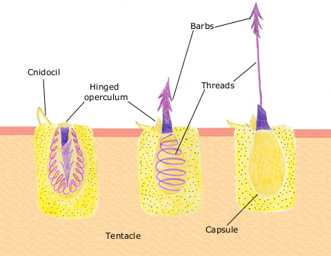
English: The diagram above shows the anatomy of a nematocyst cell and its “firing” sequence, from left to right. On the far left is a nematocyst inside its cellular capsule. The cell’s thread is coiled under pressure and wrapped around a stinging barb. When potential prey makes contact with the tentacles of a polyp, the nematocyst cell is stimulated. This causes a flap of tissue covering the nematocyst—the operculum—to fly open. The middle image shows the open operculum, the rapidly uncoiling thread and the emerging barb. On the far right is the fully extended cell. The barbs at the end of the nematocyst are designed to stick into the polyp’s victim and inject a poisonous liquid. When subdued, the polyp’s tentacles move the prey toward its mouth and the nematocysts recoil back into their capsules. |
| Fecha | 11 de abril de 2007 (fecha original de carga) |
| Fuente | Transferido desde en.wikipedia a Commons. |
| Autor | The original uploader was Spaully de Wikipedia en inglés. |
Licencia
Este archivo esta bajo la licencia Creative Commons Compartir-Igual 1.0.
Creative Commons ha retirado esta herramienta legal y no recomienda que se aplique a ninguna obra.
|
Esta imagen es de dominio público porque contiene material que vino originalmente de la Administración Nacional Oceánica y Atmosférica de los Estados Unidos de América, recibidas o hechas en el curso de las funciones oficiales de un empleado.
|
Registro original de carga
Aquí se muestra la página de descripción original. Los siguientes nombres de usuario se refieren a en.wikipedia.
- 2007-04-11 17:10 Spaully 480×371×8 (194868 bytes) Modified from: http://www.oceanservice.noaa.gov/education/kits/corals/media/supp_coral01b.html {{Information |Description=Nematocyst discharge process. |Source= Modified from [http://www.oceanservice.noaa.gov/education/kits/corals/media/supp_coral01b.html
Historial del archivo
Haz clic sobre una fecha y hora para ver el archivo tal como apareció en ese momento.
| Fecha y hora | Miniatura | Dimensiones | Usuario | Comentario | |
|---|---|---|---|---|---|
| actual | 17:29 13 oct 2007 | | 480 × 371 (190 kB) | Alison | {{Information |Description===Description== The diagram above shows the anatomy of a nematocyst cell and its “firing” sequence, from left to right. On the far left is a nematocyst inside its cellular capsule. The cell’s thread is coiled under pressur |
Usos del archivo
No hay páginas que enlacen a este archivo.
Uso global del archivo
Las wikis siguientes utilizan este archivo:
- Uso en ca.wikipedia.org
- Uso en ceb.wikipedia.org
- Uso en en.wikipedia.org
- Uso en fr.wikipedia.org
- Uso en hr.wikipedia.org
- Uso en id.wikipedia.org
- Uso en it.wikibooks.org
- Uso en ja.wikipedia.org
- Uso en lv.wikipedia.org
- Uso en ms.wikipedia.org
- Uso en my.wikipedia.org
- Uso en pa.wikipedia.org
- Uso en pt.wikipedia.org
- Uso en simple.wikipedia.org
- Uso en sv.wikipedia.org
- Uso en te.wikipedia.org
- Uso en th.wikipedia.org
- Uso en vi.wikipedia.org