
Archivo:Fusiform face area face recognition.jpg
No se dispone de una resolución más alta.
Fusiform_face_area_face_recognition.jpg (475 × 503 píxeles; tamaño de archivo: 95 kB; tipo MIME: image/jpeg)
Resumen
| Descripción |
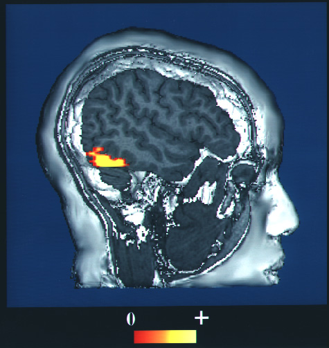
English: This is a computer-enhanced fMRI scan of a person who has been asked to look at faces. The image shows increased blood flow in the part of the visual cortex that recognizes faces.
日本語: fMRI。顔を見るように言われた人の脳内で、視覚皮質の顔情報を処理する部位で、血流増加が起きている、という画像。 |
| Fecha | upload to commons at 2009-10-27 |
| Fuente | https://www.nlm.nih.gov/hmd/emotions/frontiers.html (archive.org) |
| Autor | NIH |
| Permiso (Reutilización de este archivo) |
Public domain US government |
This image is a work of the National Institutes of Health, part of the United States Department of Health and Human Services, taken or made as part of an employee's official duties. As a work of the U.S. federal government, the image is in the public domain.
 |
||
| Esta obra ha sido identificada como libre de las restricciones conocidas en virtud del derecho de autor, incluyendo todos los derechos conexos. | ||
Historial del archivo
Haz clic sobre una fecha y hora para ver el archivo tal como apareció en ese momento.
| Fecha y hora | Miniatura | Dimensiones | Usuario | Comentario | |
|---|---|---|---|---|---|
| actual | 03:38 27 oct 2009 | | 475 × 503 (95 kB) | Was a bee | == {{int:filedesc}} == {{Information |Description= '''en:''' This is a computer-enhanced fMRI scan of a person who has been asked to look at faces. The image shows increased blood flow in the part of the visual cortex that recognizes faces. '''ja:'''fM |
Usos del archivo
La siguiente página usa este archivo:
Uso global del archivo
Las wikis siguientes utilizan este archivo:
- Uso en en.wikipedia.org
- Uso en fa.wikipedia.org
- Uso en fr.wikipedia.org
- Uso en he.wikipedia.org
- Uso en hr.wikipedia.org
- Uso en hy.wikipedia.org
- Uso en it.wikipedia.org
- Uso en ja.wikipedia.org
- Uso en ko.wikipedia.org
- Uso en ml.wikipedia.org
- Uso en nl.wikipedia.org
- Uso en pl.wikipedia.org
- Uso en simple.wikipedia.org
- Uso en tr.wikipedia.org